The National Research Foundation of Korea said Tuesday that a joint research team has found a possible way to distinguish cancer from healthy lung tissues in fluorescent contrast agents used for testing liver function.
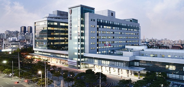
The joint research team of the Korea University Guro Hospital and the Korea Advanced Institute of Science and Technology recently developed a technique to accurately find lung cancer lesions by inhaling fluorescent contrast agent and minimize resection.

Medical professionals have previously traced lung cancer by intravenously injecting indocyanine green, a fluorescent dye used in diagnostic tests, as it tended to accumulate in cancer tissues. However, the detection required overdose and induce systemic side effects.
The research team confirmed that the indocyanine green got to the lung more efficiently by inhaling instead of injecting through the vein and saw the agent only reached healthy alveolus.
Researchers verified the inhaling method's efficiency by observing the interface of human lung cancer tissues and animal models with a fluorescence microscope.
According to the researchers, the inhaling method can reduce the necessary amount of indocyanine green by 20 times and minimize effects on other organs. This is because the agent travels to the lung focused rather than moving throughout the body via blood vessels.
However, a follow-up study on the toxicity of inhaling indocyanine green is required for the actual clinical application. Researchers plan to conduct related studies further.
The study results were published in the Journal of the American Medical Association Surgery on June 24.

