A team of researchers in Korea has published a study proving the clinical feasibility of using a miniaturized confocal laser endomicroscopy to identify cancerous tissues accurately during brain surgery.
Failing to remove brain tumors completely could cause recurrence. However, removing healthy brain tissues can cause fatal neurological disorders. Surgeons’ naked eyes are often limited so they often use surgical brain navigation devices or fluorescent dyes. But this too proves difficult to identify tumor cells precisely.
Accordingly, a research team led by Professor Kang Shin-hyuk of Neurosurgery at Korea University Anam Hospital and domestic start-up, VPIX Medical, developed a real-time digital tissue biopsy platform technology called cCELL to solve this problem.
They confirmed that cCeLL can accurately diagnose not only the distinction between normal tissue and tumor tissue but also various brain tumor cells and tissues in real-time.
The research team said cCeLL improves the accuracy and reduces the time of surgery as it can distinguish normal brain tissue from tumor tissues within three minutes, minimize damage to normal tissue, and accurately excise tumor tissues.
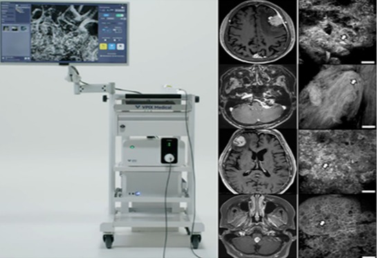
The ultra-small confocal laser endomicroscopy (CLE) is equipped with a high resolution that can view cells in the body. Unlike general microscopes, microstructures of cells and surrounding tissues can be intuitively observed, magnified, and reconstructed into two-dimensional and three-dimensional images. This information can then be transmitted to picture archiving systems (PACS) in hospitals and tissues can also be recorded to enable remote pathology diagnosis between neurosurgeons and pathologists during surgery.
The researchers plan to continue developing the device so that cCeLL can be applied to other cancerous organs such as the kidney, stomach, and prostate.
"We developed this device after years of trial and error between doctors and engineers and expect cCELL to be internationally competitive because it is now entering medical institutions around the world,” Professor Kang said.
"It is a very encouraging result for the medical device industry that this fast and accurate diagnosis of brain tumors has been verified with domestic technology,” VPix Medical CEO Hwang Kyung-min said. "As we have a number of exclusive patents, we will definitely commercialize them as products and try to lead the global medical market."