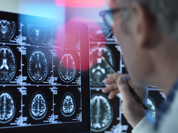
A recent study conducted by a joint research team from the Institute for Basic Science (IBS), KAIST, and Severance Hospital has successfully visualized reactive astrocytes during neuronal hypometabolism, presenting a way for early diagnosis of Alzheimer's disease.

Alzheimer's disease is typically associated with inflammatory responses, such as reactive astrogliosis, which leads to alterations in the size and function of astrocytes in response to injury, disease, or infection.
While astrocytes typically help maintain brain homeostasis, in brain diseases such as Alzheimer's disease, an increase in the number and size of astrocytes is often accompanied by various functional changes.
The researchers had previously found that the induction of reactive astrocytes is associated with the presence of the enzyme monoamine oxidase B (MAO-B), a protein located in the mitochondria of cells that facilitates the breakdown of monoamines. MAO-B is also known to play a significant role in the metabolism of neurotransmitters in the nervous system.
Despite the clinical significance of reactive astrogliosis, there is currently no neuroimaging technique available to effectively visualize them at a clinical level for the purposes of observation and diagnosis.
This study demonstrated that positron emission tomography (PET) imaging, using carbon 11-acetate and fluorodeoxyglucose, can be used to visualize reactive astrocytes and the subsequent reduction in glucose metabolism in neurons in Alzheimer’s disease.
In animal studies, the researchers found that reactive astrocytes activate acetic acid metabolism in reactive astrocytes and induce inhibition of glucose metabolism in surrounding neurons. They also revealed that acetic acid promotes reactive stellate cellularization, leading to the production of putrescine and GABA, which can cause Alzheimer’s.
In particular, the excessive uptake of acetic acid in reactive astrocytes by the monocarboxylate transporter 1 (MCT1), which is specifically expressed in astrocytes, further promoted reactive astrogliosis.
Conversely, the suppression of reactive astrocytes or MCT1 expression restored acetic acid metabolism in astrocytes and glucose metabolism in surrounding neurons.
The researchers noted that although amyloid beta has been traditionally considered the primary cause of Alzheimer's disease, its efficacy in diagnosing patients has been limited, and drugs targeting amyloid beta have not been successful in treating the disease.
"The reactive astrocytes showed metabolic abnormalities that resulted in excessive uptake of acetic acid, unlike the normal state," said Professor Yun Mi-jin of the Nuclear Medicine Department at Severance Hospital. "We found that this uptake of acetic acid plays an important role in promoting intracellular inflammatory responses."
Professor Justin Lee of IBS who led the research added that the study points to the suppression of MCT1, as a new therapeutic target for Alzheimer’s disease.
The study was published in the Brain journal on Monday.
Related articles
- Dementia society skeptical about case’s name change to ‘cognitive disease’
- Neurophet AQUA scores Singapore's approval for its brain image analysis software
- Korea's microbiome industry shows robust growth despite late start, experts say
- KAIST scientists use CRISPR to develop TCR-T immunotherapy for solid cancers

