Korean researchers have developed the world's first technology for high-speed three-dimensional imaging of blood cells in blood vessels without contrast agents.
The Korea Advanced Institute of Science and Technology (KAIST) said Wednesday that a research team led by Professor Oh Wang-yuhl of the Department of Mechanical Engineering and the KI for Health Science Institute has developed the technology. The new technology can take 1,450 images per second of blood cells flowing in various complex blood vessels without using substances such as fluorescent contrast agents.
Currently, laser scanning confocal and multiphoton microscopy techniques are used for high-resolution blood flow imaging. However, red blood cells or plasma, fluorescently stained with a contrast agent, must be tracked with a specially designed scanning method. This method has limited temporal resolution and a small number of vessels that can be analyzed for hemodynamics. Optical coherence tomography (OCT)--based techniques are also limited, as they can only analyze blood flow velocity indirectly.
The researchers developed an image processing method designed to exploit the characteristics of flowing blood cells and succeeded in imaging only flowing blood cells from microscopic images. They also used spatially uncorrelated illumination to overcome the problem of speckle noise, which obscures blood cells. The camera's high light intensity makes it possible to image blood cells flowing deep in the body at high speeds.
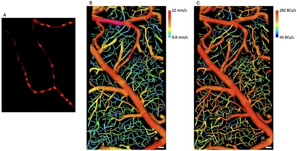
"Blood flow velocity in various blood vessels and the number of blood cells flowing per unit of time are very important information for biomedical research and have been the focus of much research for a long time," Professor Oh said. "The newly developed technology can directly image only blood cells flowing in various blood vessels at high speed without injecting any substances, including fluorescent contrast agents, into the body."
Not only is it very convenient to use in the field, but it also provides accurate hemodynamic information immediately, which will be very useful in research," Oh added.
The paper, "Direct Blood Cell Flow Imaging in Microvascular Networks," for which KAIST student Kim Gyoung-hwan and Dr. Park Hyun-sang are co-lead authors, was published in the October issue of “Small,” an international journal for convergence research.