A technology has been developed to visualize micro RNAs (miRNAs) in cells in high-resolution images.

Experts expect it to enable early diagnosis and prediction of various diseases, including cancer, myelopathy, diabetic kidney disease, and Alzheimer's disease, in body fluids such as blood.
Gangnam Severance Hospital said Wednesday that its research team led by Professor Park Jeong-yoon of the Department of Neurosurgery has developed a technology to visualize micro RNA (miRNA) present in cells in high-resolution images.
Currently, tissue biopsy is the standard method used to diagnose and monitor diseases. It involves removing tissue through surgery or a procedure, which requires invasive access to the tissue and is burdensome for the patient. Sometimes, biopsies are impossible, depending on the disease and patient's circumstances.
Also, there is heterogeneity in the biological characteristics of the same tumor depending on where it is collected.
Liquid biopsy is emerging as an alternative to overcome these limitations of tissue biopsy. By utilizing body fluids, such as blood, saliva, or cerebrospinal fluid, it is possible to identify DNA alterations by simply collecting a fluid sample.
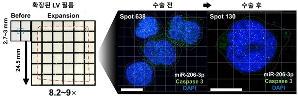
The research team developed a hydrogel film, the Liquid View (LV) film, which has the property of expanding as it absorbs liquid (distilled water). When liquid samples containing body fluids, such as blood, spinal fluid, and saliva, were absorbed into the LV film and the incubation and hardening process was repeated, the film expanded up to nine times while remaining as thin as 1.25 millimeters thick.
That enables high antibody penetration, allowing for clear observation of cell nuclei and cytoskeletal structures without antibody diffusion.

The researchers applied the LV film to body fluids (blood, urine) from patients with Alzheimer's disease (AD), spinal cord injury (SCI), and diabetic nephropathy (DN) to analyze miRNA ultra-high-resolution imaging. The results showed that ultra-high-resolution imaging detection and quantification of miR-206-3p in peripheral blood mononuclear cells (PBMCs) from AD and spinal cord injury patients was possible.
Furthermore, LV films were used to analyze the differences in miRNA expression in the blood of myelopathy patients before and after surgery. The results showed that the expression of certain miRNAs (miR-206-3p, miR-181b-5p, miR-26b, miR-27b-3p) was increased in the blood of myelopathy patients before surgery.
The researchers found a decrease in miR-206-3p and an increase in miR-206-5p after surgery and confirmed the same pattern in animal model blood.
The results of the experiments using LV films are consistent with other previous studies, demonstrating that LV films are suitable for detecting small amounts of miRNAs in cells, according to the hospital.
In particular, it is very useful for controlling the scalability of hydrogels and precise imaging quantification of small molecules, including miRNA, which is expected to be suitable for long-term experiments, such as library construction through large-scale screening studies.
“We expect this research to advance the practical use of liquid biopsy technology,” Professor Park said. “LV film enables high-resolution visualization of miRNAs in cells. The ability to detect miRNAs with higher accuracy and sensitivity will help us to detect subtle changes, as well as diagnose, prognosis, and predict diseases.”
The paper, "Customized Hydrogel Films for MicroRNA Super-Resolution Imaging in Liquid Biopsies,” was published in Advanced Healthcare Materials, an international journal specializing in nanomedicine and biomaterials.