A study conducted by Severance Hospital has shown that optical coherence tomography (OCT)-guided intervention is remarkably effective in treating complex coronary artery stenosis.

Coronary artery stenosis occurs when the coronary arteries, which supply blood to the heart, become narrowed due to thrombus formation, leading to impaired blood flow. This condition can result in diseases such as angina. The stenosis often manifests in a variety of complex forms, including chronic total occlusions, bifurcation lesions, left main disease, and calcified lesions; collectively termed "complex coronary artery stenosis."
Traditionally, percutaneous coronary intervention (PCI), which involves injecting contrast agents and inserting stents, has been the standard treatment for coronary artery stenosis.
However, when applied to complex stenosis cases, PCI presents challenges, including increased difficulty in stent placement, lower implantation success rates, and a higher risk of complications, such as vessel dissection.
To address these challenges, the research team -- led by Professors Kim Byeong-keuk, Hong Sung-jin, and Lee Seung-jun from the Cardiology Department at Severance Cardiovascular Hospital, along with Professor Emeritus Jang Yang-soo from Yonsei University -- focused on OCT technology.
By capturing 3D images of blood vessels, OCT enables precise anatomical assessment and aids in developing tailored treatment plans for complex stenosis.
The team analyzed the outcomes of 1,604 patients with complex coronary artery stenosis treated at over 20 institutions across Korea from January 2019 through September 2022.
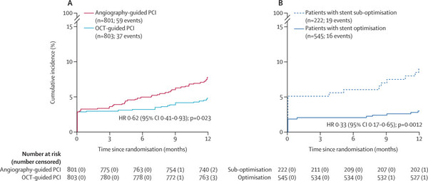
The results showed that the incidence rate of adverse events such as cardiovascular death, myocardial infarction, stent thrombosis, and ischemia-induced vascular revascularization was 7.4 percent in the group that underwent conventional PCI procedures based on contrast agent administration (n=801).
In contrast, the rate of complications was 4.6 percent in the OCT-guided intervention group (n=803) -- which was approximately 38 percent lower than the PCI group.
The team attributed the improved outcomes to better stent optimization achieved through OCT guidance. The 3D imaging provided by OCT allowed for more precise stent sizing and positioning, as well as reduced damage to the vessel walls during the procedure.
Indeed, surgeons performing OCT-guided interventions could better determine the appropriate stent size and placement for each patient.
Post-procedure, OCT also facilitated additional treatments, such as balloon dilatation, to further improve stent optimization.
“OCT-guided intervention offers 3D imaging of the coronary arteries, significantly aiding in the development of personalized treatment plans,” Professor Kim said. “This study is the first to demonstrate the improved outcomes of OCT-based interventions for complex coronary artery disease, and it is expected to influence future treatment guidelines.”
The results of the research were published in The Lancet.
Related articles
- 3rd-gen TKI proven effective in treatment-resistant EGFR-mutant lung cancer with brain metastasis: study
- Severance Hospital develops method to verify retinal function
- Severance Hospital tops 2024 Brand Customer Satisfaction Index for tertiary hospitals
- Filipino boy gets successful heart surgery at Severance Hospital
- Severance Hospital conducts cochlear implant surgeries for children in Cambodia
- Blood vessels found to be key to cardiovascular disease: study
- Severance Hospital develops new spray treatment for allergic respiratory diseases
- Altering dendritic spine shapes in brain neurons can regulate gambling addiction: study