Researchers at the Institute for Basic Science (IBS) have discovered a way to map the cerebral cortex activity of living mice to study brain circuits using a beam projector as optogenetic stimulation combined with functional magnetic resonance imaging (fMRI).
fMRI scans the activity of the entire brain to identify changes in the entire brain caused by activity in certain optogenetically induced brain regions. Optogenetics is a biological technique to control the activity of neurons or other cell types by using light as a stimulant.
This technique is widely used to study the causal relationship between a single brain region and the entire brain circuit.
This is usually accomplished by inserting optical fibers into the target area of the brain through invasive surgery and transmitting light through them to stimulate cells. However, the conventional method could cause unexpected damage to the brain, and only a limited number of optical fibers can be inserted into one experimental animal.
Consequently, a large number of experimental animals are needed to study numerous brain areas. In addition, it is difficult to control differences between experimental subjects to study connectivity differences between brain regions.
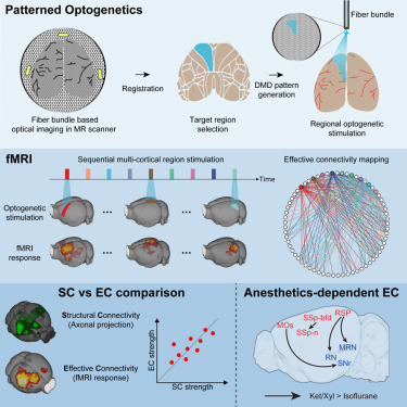
The research team, led by Professor Choi Myung-hwan of Seoul National University’s School of Biological Sciences, used a beam projector equipped with a high-power optogenetic stimulation laser to direct light directly throughout the cerebral cortex.
They used the thinned-skull cranial window technique to penetrate the mouse's skull by drilling a small hole in the brain and treating the skull with chemicals. In this way, the skull is not completely removed, thus minimizing side effects and allowing the brain to be investigated in a natural state.
In particular, the light stimulation can be applied to a wide surface of the brain so that stimulation can be delivered to the desired brain area rather than one fixed area.
Using this technique, the researchers anesthetized mice with isoflurane and ketamine-xylazine and found that isoflurane selectively weakens the connectivity between certain regions of the cerebral cortex and the middle region.
"This study demonstrates that fMRI combined with flexible optogenetic stimuli can measure dynamic changes in brain circuits induced by certain brain states," said Kim Seong-hoon, the first author of the paper. "It also presented a new method of extracting brain circuits related to specific brain conditions, which is expected to play an important role in revealing the neurophysiological mechanisms of brain dysfunction caused by brain diseases and drugs."
The study was published online in the international journal, Neuron.
Related articles
- GemVax’s Alzheimer's drug GV1001 begins phase 2 clinical trials in Spain
- Coreline Soft’s brain hemorrhage-detecting software wins approval in Korea
- ‘World's 1st' fetal brain stem cell transplant effective in Parkinson's disease: study
- Treatment mechanism found for autistic epilepsy patients with Ank2 deficiency
- IBS develops brain mapping to predict tendency to focus on negative thoughts, level of depression