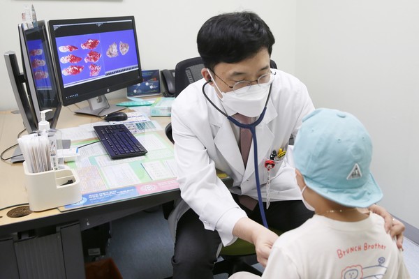
Asan Medical Center said that it has improved the survival rate of pediatric patients with liver cancer, from 60 to more than 90 percent, by applying various surgery techniques.

"Hepatoblastoma in children is a malignant tumor of the liver in children, accounting for more than 95 percent of liver cancer in children under five years old," the hospital said. "Although hospitals use chemotherapy to reduce the size of the tumor and perform surgery for complete resection, it is difficult to remove all the tumors by surgery, and the prognosis is poor if the tumors have already metastasized to other organs."
To raise the survival rate of these patients, the AMC team subdivided the intensity of chemotherapy for pediatric hepatoblastoma patients and introduced an imaging technology that uses fluorescent dyes to confirm the extent of the tumor.
The researchers increased the survival rate of pediatric hepatoblastoma patients to over 90 percent. Professors Im Ho-joon, Koh Kyung-nam, Kim Hye-ri of the Department of Pediatric Oncology and Hematology, and Kim Dae-yeon and Namgoong Jung-man of the Department of Pediatric Surgery led the research.
It could also minimize side effects by administering weak chemotherapy to pediatric liver cancer patients, who could receive operation easily, and high-intensity chemotherapy for patients with multiple tumors or metastases so that they could receive surgery.
They also injected the indocyanine green, a fluorescent that dyes normal hepatocytes, liver cancer, and hepatoblast carcinoma cells green, into the body of patients and used imaging technology with a near-infrared camera.
They used the method as normal hepatocytes excrete indocyanine green through the biliary tract. Still, hepatocellular carcinoma and hepatoblast carcinoma cells do not excrete indocyanine green, and the fluorescent signal remains even after two days.
"This fluorescence imaging system distinguishes between the surface of the liver and tumors near the resection section, and can detect even small tumors on the surface of the liver not detected by computed tomography (CT) or magnetic resonance imaging (MRI), enabling much more accurate and safe liver resection and liver transplantation," the team said.
"I think that the improvement in the survival rate of pediatric liver cancer patients is the result of considering and implementing the optimal treatment method according to each patient's condition," Professor Koh said. "We also believe that close cooperation between the pediatric oncology and hematology department and the pediatric surgeon department greatly contributed to the improvement of treatment outcomes."
Professor Namgoong also said, "The pediatric solid cancer team is collaborating with the pediatrics department to treat patients with various solid cancers, such as neuroblastoma and sarcoma, and hepatoblastoma."
In the case of hepatoblastoma, unless a liver transplant is unavoidable, the team avoids liver transplantation as much as possible and used a multi-stage liver resection to reduce the burden on patients who are afraid of transplantation, the professor added.
Cancer Medicine has published the study.

